A large amount of fish is spoilt each year from post-catch to consumption. There is a growing interest in applying essential oils (EOs) as bio-preservatives to control the spoilage of chilled-stored fish. However, piles of information on the EOs’ effects on chilled stored fish cannot provide a clear guide toward the appropriate application of EOs to restrain various spoilage microorganisms. In our recent article, we reviewed and meta-analyzed 180 scientific articles working with chilled stored fish to corroborate the promises of EOs in the chilled-stored fish industry.
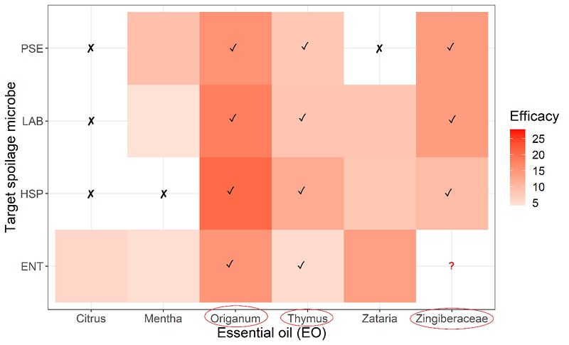
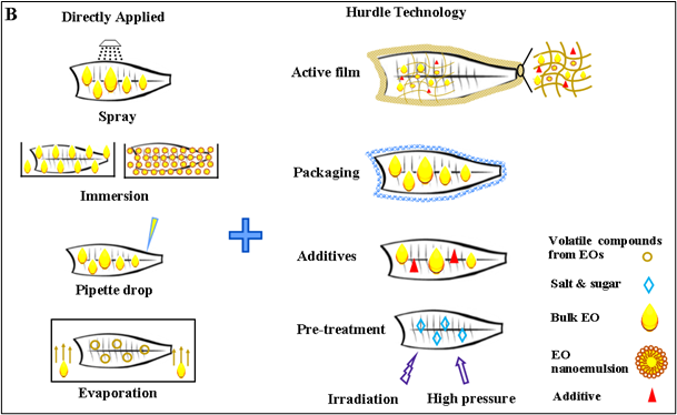
We identified six EOs with extraordinary total microflora reduction potential for raw fish – citrus, mentha, origanum, thymus, zataria, and Zingiberaceae. Not all EOs can suppress all groups of specific microbes. Only origanum, Zingiberaceae, and thymus have complete-spectrum efficacy. Noticeably, they should be applied by the correct methods, e.g., hurdle technology, active film nanoemulsion, special packaging. We also exposed the concertation effects of EOs on fish odour and flavour. It is showed that the low level of most EOs imparts little interference on the natural odour of fresh fish. Sensory deterioration in EO treated fish flesh is much slower than normal refrigerated ones. Thus, selected EOs at mild concentrations with the right application method can promote chilled-stored fish' safety, sensory and shelf-life agendas.
The study provides clear-cut strategical support to good EO’s application in chilled stored fish based on metadata analysis.
Detailed information can be found in the original paper Hao, R., Roy K., Pan, J., Shah, B.R., Mraz, J. 2021. Critical review on the use of essential oils against spoilage in chilled stored fish: A quantitative meta-analyses. Trends in Food Science & Technology 111: 175–190.
Heatmap of the anti-microbial efficacy of top 6 essential oils with extraordinary total viable count reduction potential against specific spoilage microbes
Commonly used application methods of essential oils in chill-stored fish
Vertebrates are characterized by formation of outer surface by a sheath of cells called ectoderm, while the inner lining of the primitive gut is made from endoderm. These two layers get together in the anterior part of digestive tract, so called farynx. The team of Rober Černý with Martin Minařík as the first author (Charles University in Prague, Faculty of Science, Czech Republic) came with findings, which rewrite the present opinions of developmental and evolutionary biology. The basal ray-finned fishes such as bichirs, gars and sturgeons, sometimes considered living fossils, have a facial part of head composed of endodermal cells of a pre-oral part of the gut, which is very archaic characteristic expected only in ancestors of vertebrates.
Our Faculty of Fisheries and Protection of Waters supplemented the study with a representative of sturgeons and with two co-author positions (Martin Pšenička and David Gela) and two other colleagues mentioned in acknowledgement (Marek Rodina and Martin Kahanec) became inscribed in the prestigious journal Nature.
For more info see a press release here: https://www.natur.cuni.cz/fakulta/aktuality/stara-tajemstvi-nove-hlavy-obratlovcu or the manuscript on the official web page of Nature: http://www.nature.com/nature/journal/v547/n7662/full/nature23008.html, or a publicly accessible view-only version: http://rdcu.be/tXoa
TV interview on ČT24 starts at 15:34 minute:
http://www.ceskatelevize.cz/ivysilani/10101491767-studio-ct24/217411058290719
Full reference: Minarik, M., Stundl, J., Fabian, P., Jandzik, D., Metscher, B.D., Psenicka, M., Gela, D., Osorio-Pérez, A., Arias-Rodriguez, L., Horácek, I., Cerny, R. Pre-oral gut contributes to facial structures in non-teleost fishes. Nature, 547: 209-212. doi:10.1038/nature23008 (IF=40.137)
-----------------------------------------------------------------------------------------
Picture legend: SEM images of sturgeon head with endodermal contribution pseudocoloured yellow. Antero-ventral views, 10 days post hatching; mb, medial barbel; lb, lateral barbel; ao, ampullary organs; rb, rostrum; llo, lateral line organs; ll, lower lip.
3-D reconstruction of a live cell using the new technology Credit: Dr Renata Rychtáriková
Looking at live cells under a microscope is a difficult task. The size and shape of samples limit the methods biologists can use to look at their materials, and even what they can examine. But a new algorithm could help researchers get a better look at living cells, with research applications from cancer treatments to IVF.
“We live in the nano-era,” says Dr Renata Rychtáriková, the paper's lead author and a researcher at the University of South Bohemia, in the Czech Republic. But, she notes, things at a nano scale are hard to see. “The majority of the nano-world escapes observation at the moment, and our information about it is only indirect.”
Current sample-studying methods, such as Scanning Electron Microscopy (SEM), only reveal what is happening on the surface. Samples for other methods, like transmission electron microscopy, must have a thickness between 10 and 200 nm, while a typical mammalian cell has a thickness of 3-5 μm.
To combat this, Rychtáriková and colleagues developed a way to use bright-field optical microscopes to delve deeper into objects. Their algorithm captures several images to build a 3-D map of a cell. They have published their results in the journal Ultramicroscopy.
The algorithm works by comparing two images taken at slightly different positions. It looks for parts of the cell that respond best to the light, and seeks out ones that are moving. By doing so, it helps shine a light on detailed cellular structures more effectively than previous methods. The result is a virtual 3-D model of the cell.
“The research was inspired by the biology of living mammalian cells, which is fundamental for cancer healing, organ replacement, implantology, neurology, embryology and IVF,” explains Rychtáriková. “There has been great interest from the medical community since the start of the development of our method,” she adds. And the research is attracting interest from several other sources as well.
“After the biologists, the material scientists interested in nanoprinting came forward,” says Rychtáriková. Because the algorithm allows researchers to use thicker samples, and because it only requires a common microscope, many scientists are hoping to start using it. “The interest from the application sphere far exceeds our time possibilities,” she adds.
The article was assigned by Elsevier among top 30 papers in the field of Physics for 2017.
Article details:
Rychtáriková, R., Náhlík, T., Shi, K., Malakhova, D., Macháček, P., Smaha, R., Urban, J., Štys, D. 2017. Super-resolved 3-D imaging of live cells’ organelles from bright-field photon transmission micrographs. Ultramicroscopy 179: 1-14. (IF 2015 = 2.874)
Caviar is a high cost delicacy consisting of salt-cured fish-eggs of the sturgeon family. Natural populations of sturgeon have declined during the past century through poaching for caviar, water pollution, and habitat degradation, making them currently the world’s most endangered group of species. It has led to the development of sturgeon culture, originally for reintroduction, but more recently for caviar production. “Besides pure species, less priced caviar is also produced by fertile sturgeon hybrids. Among those, bester (hybrid between beluga Huso huso female and sterlet Acipenser ruthenus male) is the most widely used.
Caviar from hybrids is almost impossible to identify from pure species caviar without reliable DNA-based technique. Similarly, identification of sturgeon species is very complicated and requires molecular genetic techniques” says Milos Havelka, who works on sturgeon research at Faculty of Fisheries and Protection of Waters, University of South Bohemia.
Researchers from University of South Bohemia in cooperation with Hokkaido University in Japan developed new method for identification of beluga sturgeon and its caviar. Beluga caviar is the most costly in the trade. Less valuable roe from other species or hybrids is sometimes fraudulently sold as beluga caviar. “Using modern methods of molecular genetics, we identified species-specific variants in genome of beluga and sterlet sturgeons. Taking advantage of these variants, we have developed simple method allowing identification of beluga, sterlet and their interspecific bester hybrid. Importantly, the tool works for caviar and requires only one roe for analyses” adds M. Havelka.
Developed tool should contribute to better, more reliable, regulation and control of global trade of high value sturgeon products as well as to their management and conservation. The protocol is straightforward and thus can be easily implemented across laboratories.
Detailed info is available in original article: Havelka, M., Fujimoto, T., Hagihara, S., Adachi, S., Arai, K. 2017. Nuclear DNA markers for identification of Beluga and Sterlet sturgeons and their interspecific Bester hybrid. Scientific Reports. Doi: 10.1038/s41598-017-01768-3
Figure 1. Caviar sample ready to be analysed.
Figure 2. Schematic workflow of caviar sample analysis using developed method.
Global research shows that treated municipal wastewaters still contain some xenobiotics. Progestins (also known as progestogens) are an important group of such compounds. They are contained e.g. in hormonal contraception and in other hormonal preparations what predisposes them to have a broad therapeutic use. Thus, progestins occur in municipal wastewaters and their concentrations rather tend to increase. Progestins can pass through municipal wastewater treatment plants unchanged and then contaminate surface waters. Indeed, various studies have shown their presence in surface waters worldwide. In some cases, progestins were found in surface waters at levels that exceed the concentrations inducing adverse effects on aquatic organisms under laboratory conditions. Despite that there are only few pieces of information on this topic, the risk posed by progestins for the aquatic environment should not be underestimated. Given that progestins mimic natural hormones, their uncontrolled entry into a body can seriously disrupt the established hormonal balance and could result in a myriad of other consequent adverse effects.
For these reasons, scientists from the Faculty of Fisheries and Protection of Waters in Vodňany (under the University of South Bohemia in České Budějovice) aim at screening of the occurrence of progestins and the related hormonal (progestagenic) activities in Czech aquatic environment. They focus on the “risky” localities including effluents from wastewater treatment plants and the respective downstream surface waters. Along with monitoring of occurrence of progestins and hormonal activities, experiments in laboratory were carried out in order to determine progestagenic activity of these compounds. It has been revealed that progestins do not occur at such a high levels in effluents from Czech wastewater treatment plants as they do in other European and Asian countries. Nevertheless, due to continuously increasing consumption of hormonal preparations and broadening their use, this issue deserves further attention. Results of this research also indicate which compounds, out of a wide spectrum of progestins, could pose the highest risk for the aquatic environment and therefore they should get the highest priority in further testing. These include medroxyprogesteron acetate, megestrol acetate and progesterone, which have been detected most frequently at studied localities and also possess relatively strong hormonal (progestagenic) activity.
The implementation of the current research has been ongoing for three years and is financially supported by the Grant Agency of the Czech Republic (16-09709Y). Samples of wastewater have been provided by following institutions: ČEVAK, a.s.; TSST Strakonice s.r.o., BVK, a.s. a STU Bratislava.
Detailed information on obtained results can be found in publication: Šauer, P., Stará, A., Golovko, O., Valentová, O., Bořík, A., Grabic, R., Kocour Kroupová, H., 2018. Two synthetic progestins and natural progesterone are responsible for most of the progestagenic activities in municipal wastewater treatment plant effluents in the Czech and Slovak republics. Water Research 137: 64-71.