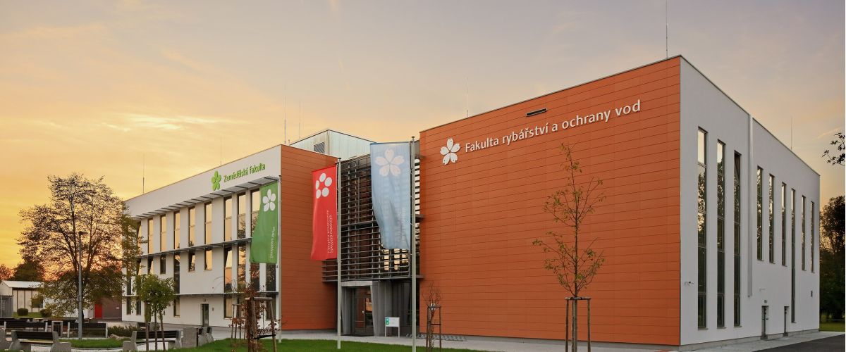
Institute of Aquaculture and Protection of Waters /IAPW/
Laboratory of Machine Vision in Aquaculture and Water Protection
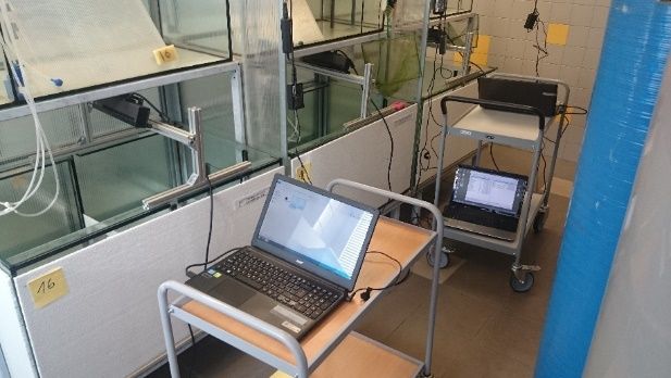
The laboratory is dedicated to applying modern technologies in aquaculture and water protection. We develop monitoring systems to track fish, crayfish, and other aquatic organisms associated with aquaculture, with an emphasis on low-cost solutions that enable real-time monitoring.
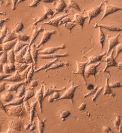
We focus not only on macroscopic monitoring using camera systems but also on microscopic observation of non-specific cellular responses to assess cytotoxicity across a wide range of samples. These analyses provide key information about the biocompatibility of materials and their potential application in human or veterinary medicine, as well as on water pollution by micropollutants, chemical compounds and their mixtures, and leachates and extracts from solid materials such as alloys, polymers, or composites.
We implement advanced data and image processing methods to analyze appearance, behavior, and to perform classification or detection tasks, utilizing convolutional neural networks.
Our main goal is to apply automation (Industry 4.0) in the aquaculture sector to support aquatic organism production while improving welfare and water protection. Automating processes through modern technologies increases competitiveness, and the use of contactless measurement and non-invasive methods contributes to improved living conditions for aquatic animals.

Key activities:
- Development of fish imaging systems
- Monitoring behavior of aquatic animals – welfare
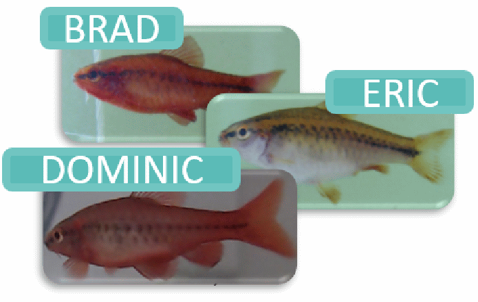
- Individual fish identification based on images
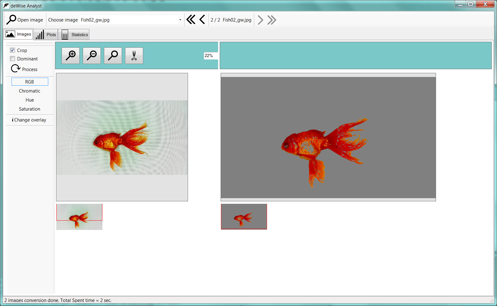
- Fish coloration analysis
- Remote water quality monitoring
- Disease detection in fish based on imaging
- In vitro toxicity and biocompatibility testing:
- Evaluation of cytotoxicity of water, micropollutants, chemical compounds, mixtures, leachates, and solid material extracts
- Providing insights on sample biocompatibility for use in material development, clinical, or animal research
We offer free collaboration through open access to our services and infrastructure.
Key projects:
Individual image based fish identification
The project aim is to substitute invasive fish tagging by non-invasive individual fish identification based on fish appearance. We already tested that fish skin patter can be used for individual identification under laboratory conditions for salmon, carp, sea bass, rainbow trout and Sumatra barb. The next step is to implement out method for under the real conditions to be useful in tanks and cages
Fish ethological study
Fish behaviour reflects the fish state. If we are able to monitor changes in behaviour or difference between behaviour of fish group, we can explore the changes of fish state. The series of projects is focused on the automatized fish behaviour parametrization of the fish influenced by nano particles, drugs or change of welfare. You can design your ethological study and we can automatically describe behaviour of your fish.
Fish skin color measurements
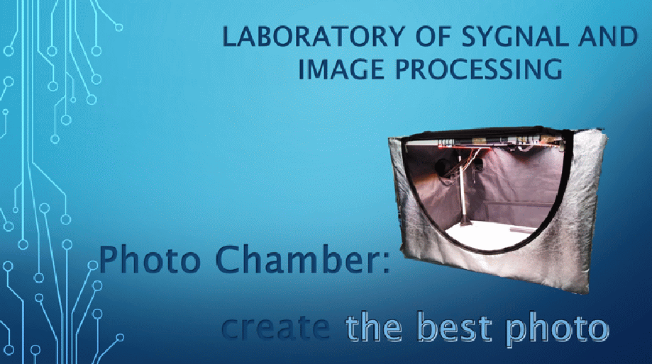
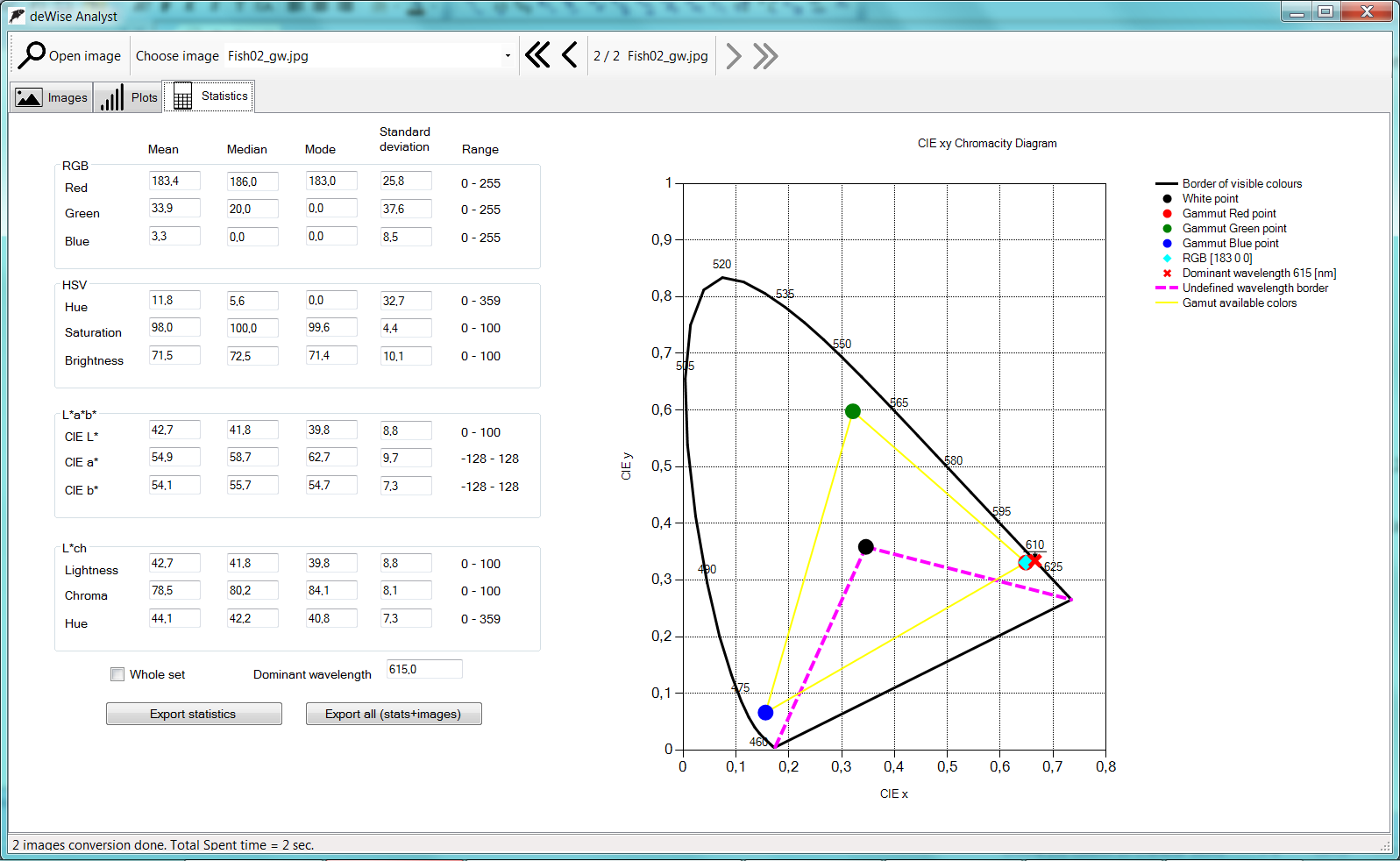
The photographical chamber offers the illumination of the fish in visible or near infrared light on standardized background. Included camera is controlled by the microcomputer and providing self-calibration of the white balance. The captured series of image could by evaluated by associated software. There are no needs for user given parameters. The basic statistical properties of the color distribution over the fish body are evaluated for wide range of color reprezentations. The advantages are the camera self-calibration, possibility of measuring wet surfaces, automatic segmentation and evaluation.
Water quality by remote sensing
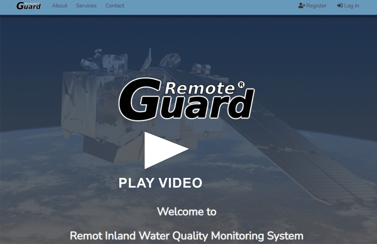
Inland water bodies (ponds, reservoirs) quality is critical for fish cultivation and for use of water bodies by the public. The standard water quality analysis is based on the water sampling and laboratory analysis which is timely and costly. We developed the system for estimation of basic (chlorophyl A) parameters from freely available satellite images. The user can select the pond and the date and the system estimates needed parameters in the case of available satellite image.
Monitoring of fish ladder
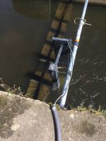
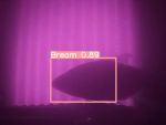
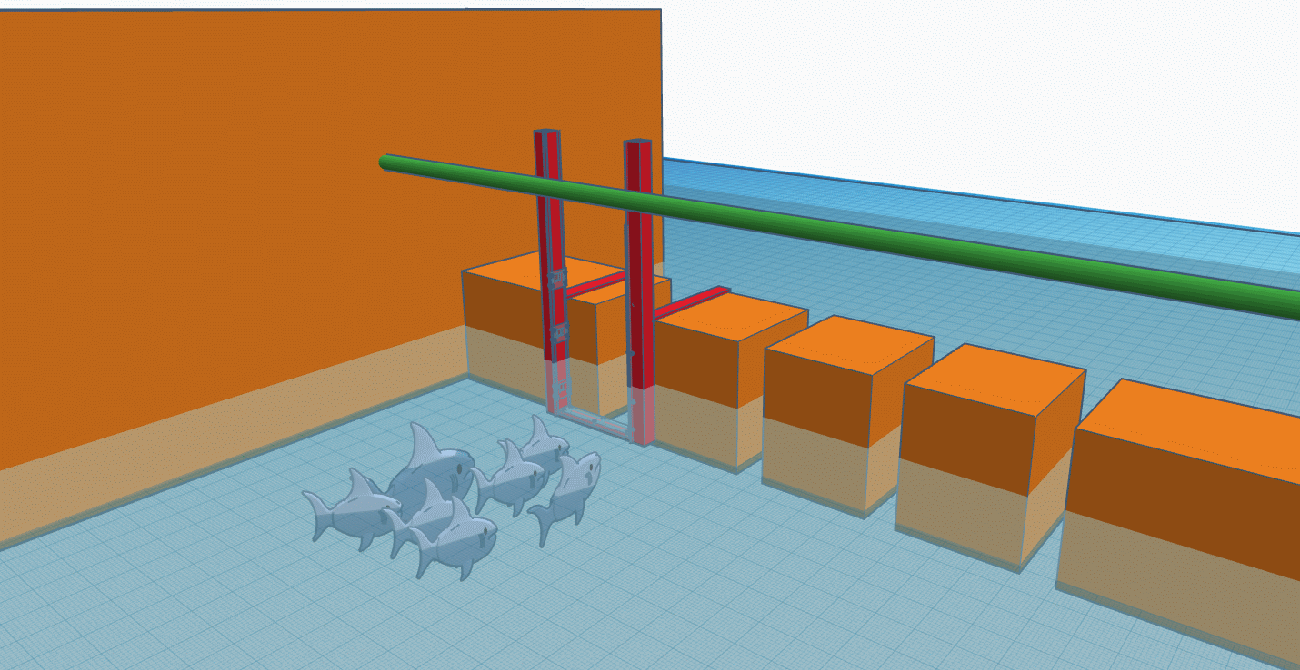
Fish ladders are structures on barriers in rivers to help fish natural migration. We developed a technology to monitor the passing fish via infrared light and camera systems. The research is focused on answering the questions, how the ladders are working, what kind and amount of fish are passing. To answer such questions we are using neural network for detection and classification.
TACR
This project focuses on developing scaffolds based on decellularized human tissue for regenerative medicine. Key steps include designing and implementing a decellularization unit using supercritical CO₂, optimizing protocols, and seeding scaffolds with mesenchymal stem cells. The resulting scaffold preserves the original 3D tissue structure and enables tissue regeneration, thereby improving healing of damaged tissues. Our laboratory evaluates the biocompatibility of these materials and provides feedback to optimize procedures for the best outcome. Once completed, the product will be registered as a medical device.
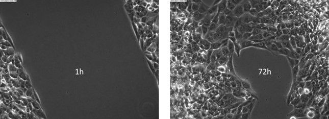
Image: Model wound healing method – one of the experimental methods we use to assess wound healing and material biocompatibility. It involves creating a controlled wound on a model (e.g., in vitro on cell cultures) and subsequently observing the effects of tested biomaterials, drugs, or cell therapies on the healing process. Parameters such as wound closure speed, cell proliferation, new tissue formation, and inflammatory response are monitored.
Automatic Live Cell Detection
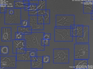
We develop methods for automated cell analysis from micro-cinematographic recordings using convolutional neural networks (CNNs). Previously manual and time-consuming evaluations have been replaced by efficient detection, accelerating data processing and improving statistical evaluation accuracy. The optimized CNN model enables detailed analysis of cell interactions with the sample, which is crucial for subsequent research phases.
Automatically detected cells using a trained CNN model
Cytotoxicity Testing of Liquid Samples
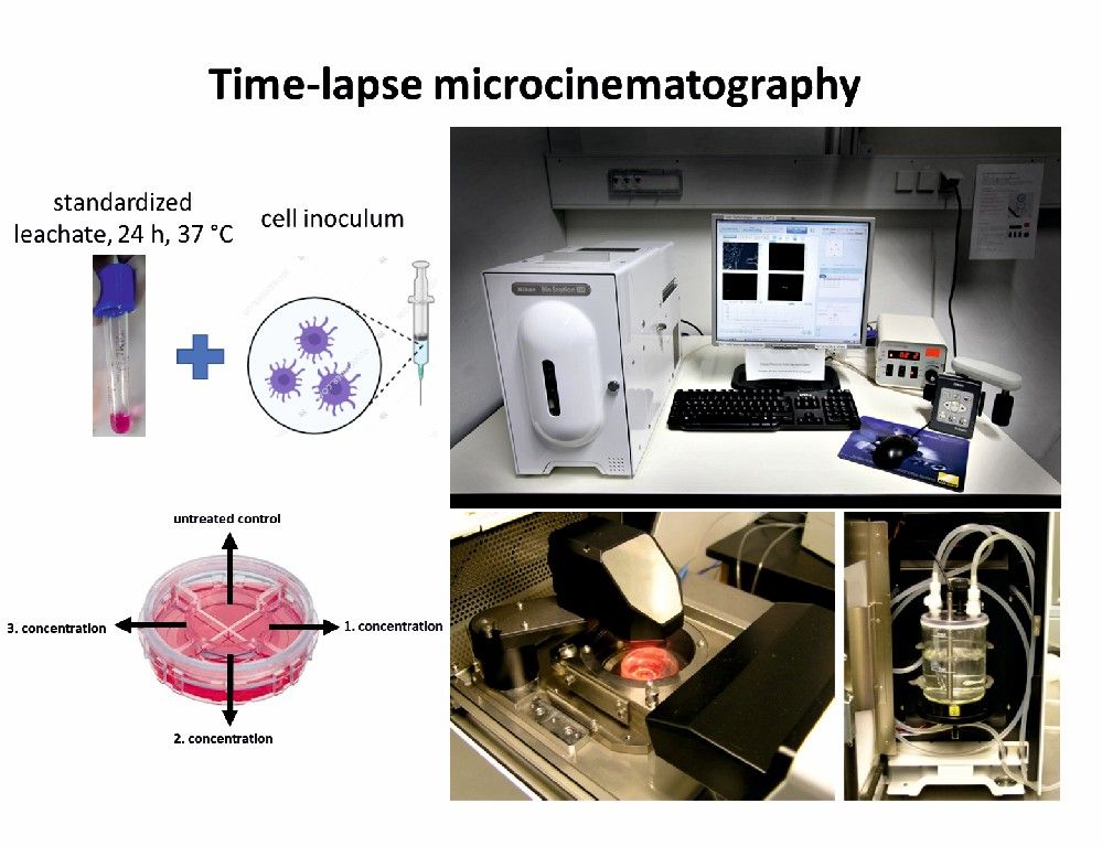
We use advanced live-cell image analysis to test the cytotoxicity of liquid samples, including leachates and solid material extracts. Time-lapse microcinematography allows long-term monitoring of cellular responses without staining, revealing even subtle morphological and growth changes. This approach offers a more precise assessment of material toxicity and biocompatibility compared to standard methods.
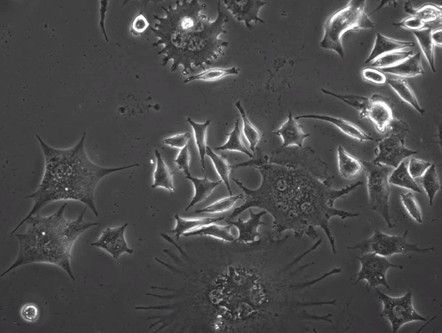
Revealed cellular anomalies
Biocompatibility Testing of Solid Samples
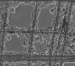
We test the biocompatibility of solid samples by monitoring the interactions of live cells with material surfaces. Using time-lapse microcinematography, we analyze cell growth, adhesion, and behavior in contact with the tested material, which allows us to assess its suitability for biomedical applications. Our methods provide detailed insights into toxicity, cellular colonization, and surface properties of materials, contributing to the development of safe and functional biomaterials.
Interaction of live cells with the surface of a titanium-based implant material (left) and polymer nanofibers (right)
Staff and contacts
-
Laboratory of Machine Vision Processing in Aquaculture and Water Protection
Institute of Aquaculture and Protection of WatersZámek 136
373 33 Nové Hrady

