
Research Institute of fish Culture and Hydrobiology /RIFCH/
About Laboratory of Germ Cells
Since its establishment in 2015, the laboratory has been interested in germ cell biology (gametes precursors) in several fish species with representatives of purely model species (zebrafish and goldfish), aquaculture species (common carp, tench, pikeperch), but also critically endangered sturgeons. Multidisciplinary approaches are used to study germ cells, including molecular analyses (gene expression, microsatellites), chromosome manipulations (induction of uniparental inheritance and polyploidy), histology, immunolabeling, flow cytometry, genome editing using the CRISPR/Cas9 system and germ stem cell transplantation with the aim of inducing chimerism and produce donor-derived offspring from surrogate parents.
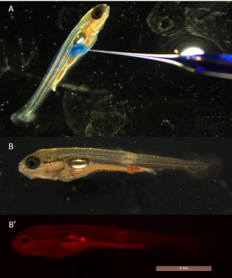
Fig. 1. Germ cell transplantation obtained from common carp into goldfish peritoneal cavity. A) Injected cell suspension is stained by methylene blue for better visibility. B) Caption of transplanted goldfish in bright field. B´) Caption taken using fluorescent channel DsRed, to visualize previously labelled germ cells using membrane dye.
The researchers of the germ cell laboratory have attained several achievements in sturgeon research, where we can mention successful nuclear transfer, elucidation of the polyspermy phenomenon, description of specification and migration of primordial germ cells, as well as freezing and subsequent transplantation of germ stem cells. One of the long-term goals of the laboratory is transplantation of germ cells from late-maturing sturgeon species into the small and short maturing sterlet host, which is expected to significantly reduce length of the reproductive cycle. In course of this long-term effort, several techniques of non-transgenic and transgenic sterilization of sterlet have been developed for this purpose.
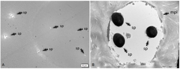
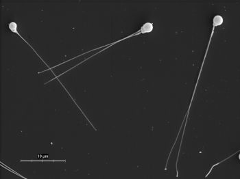
Fig. 2. Electron microscope captions on sturgeon egg having several micropyles (B), with detailed view on one micropyle with three spermatozoon, which is attributed to polyspermic fertilization resulting in atypical cleavage patterns captured on figure 3.
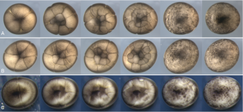
Current research topics on sturgeons in the laboratory are aimed on the molecular characterization of germ cells in juvenile individuals, which can bring new insights into sex determination and development. Cell sorting and in vitro cell culture then followed by RNA sequencing is used for this purpose. Beside sturgeon germ cell, the development of endoderm and the fate of the yolk cells in archaic fish species are studied, because their show very unique patterns.

Fig. 4. Morfology of cultured sterlet germ cells.
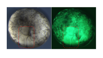
Fig. 5. Sturgeon embryo with partially blocked cleavage (area depicted by red dashed rectangle) Left brightfield picture, right picture taken on fluorescent channel.
The laboratory has two recirculation zebrafish systems for the breeding where several zebrafish transgenic lines are maintained and used for the study of germ cells through various transplantation methods, but also to study lipid metabolism via gene knockout. Zebrafish breeding colony also serves as a model for preliminary studies to be carried out in sturgeons. At the same time, laboratory members provide support to colleagues from other laboratories who conduct experiments on zebrafish. The laboratory also uses capacity of the aquarium room located at Genetic Fisheries Centre, where goldfish germline chimera producing common carp gametes are reared. Researches do not neglect less traditional and less-known fish species, such as loach, which offers unique opportunity to study sexual and asexual reproduction on the molecular level.
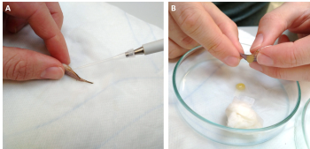
Fig.7. Gamete collection from zebrafish male (A) and female (B).
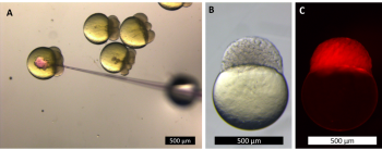
Fig. 8. Zebrafish embryos microinjection with dye (A), detectable only under fluorescent light (B, C).
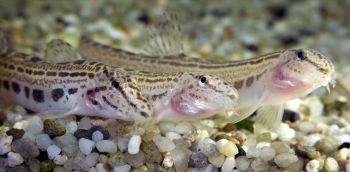
Fig. 9. Adult specimens of spined loach from laboratory colony.
The laboratory is equipped with high-end technique for visualization (fluorescent microscope and stereomicroscope with motorized drive), several stereomicroscopes with brightfield for routine micromanipulation procedures with microinjectors (pneumatic and manual) and micromanipulators (robotized and manual) including unit for piezo-assisted injection. Further equipment is flow cytometer, high capacity centrifuge (cooled), equipment for nucleic acid isolation and analysis (flow box, spectrophotometer, thermocycler, capillary and gel electrophoresis) and electroporator.
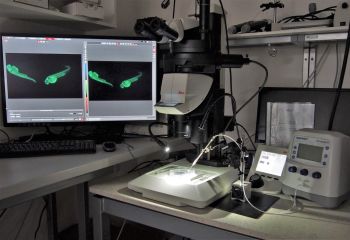
Fig. 10. Robotized fluorescent stereomicroscope with micromanipulator and microinjector for manipulation with fish embryos.

Fig. 11. Laboratory for molecular and cell analysis.
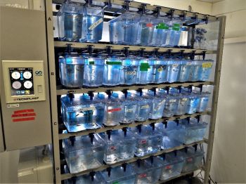
Fig. 12. Recirculation systems for zebrafish housing.
Members of the laboratory cooperate closely with several national and abroad institutions - Laboratory of Fish Genetics of the Institute of Animal Physiology and Genetics of the CAS, Laboratory of Gene Expression of the Institute of Biotechnology CAS, Laboratory of Craniofacial Evolution of the Charles University. Abroad collaborations are mainly with Laboratory of Fish Physiology and Genomics, Rennes, France and University of Ehime and Hokkaido University in Japan.
Staff and contacts
