
VEDA/LZB

A giant surrogate for zebrafish

  • Fotogalerie:
    • Foto:
    • Foto:
    • Foto:

Germ stem cells are the progenitors of gametes that can be isolated and transplanted between phylogenetically related species. The transplantation of these cells presents significant promise in reproductive biotechnology, enabling the production of gametes and offspring of the donor origin through surrogates. Zebrafish are a popular research model due to their beneficial characteristics and extensive genomic resources. Yet, their diminutive size restricts sperm yield.  In our research, we leveraged the technique of surrogate production to enhance sperm production in zebrafish by utilizing the larger Devario species, specifically the giant danio, which is three times its size. We extracted spermatogonial cells from mature zebrafish males and introduced them into sterile giant danio larvae. Half a year after the transplantation, the recipient generated sperm of zebrafish with thrice the volume and tenfold the concentration compared to zebrafish. The presented study is the first of its kind to produce zebrafish donor-derived gametes via a surrogate from different genera.

Detailed information can be found in the original article: Enhancement of zebrafish sperm production via a large body-sized surrogate with germ cell transplantation. Communications Biology 6: 412. https://doi.org/10.1038/s42003-023-04800-7

Picture: Schematic of the study.

Written by: Assoc. Prof. Martin Pšenička

Evo-devo research at FROV, the results of which have been published in prestigious journals

At the end of last year, a European project was successfully completed at our faculty, whose principal investigator was Jan Štundl, who is currently working at the California Institute of Technology in the United States and continues to work closely with the Faculty of Fisheries and Water Conservation at the University of South Bohemia.

In 2020, Jan Štundl succeeded in obtaining a prestigious Marie Skłodowska-Curie grant, which supported his postdoctoral stay in the laboratory of Prof. Marianne Bronner, a world-leading expert in neural bar and evolutionary developmental biology (evo-devo). The neural bar is a unique cell population that arises during early embryonic development. These cells subsequently migrate throughout the embryo and give rise to many different structures, including toothed jaws, various neurons or dentoskeletal elements such as shark scales or sturgeon tags. "The neural bar is only found in vertebrates and we owe it to it that we are an evolutionarily successful species," explains Jan Štundl. The cells of the neural crest are involved in almost every part of our body and also play an important role in regeneration processes.

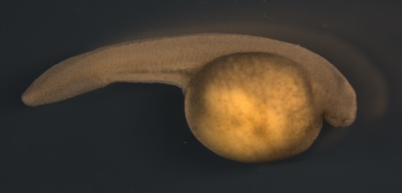
Within the Heart2019 project, Jan Štundl focused on the investigation of a specific subpopulation of the neural bar - the cardiac neural bar.  Its cells migrate to the heart during early development and play a key role in the formation of the heart muscle. This project followed pioneering research by Prof Marianne Bronner, who suggested that these cells could be responsible for heart regeneration. "Based on research on a model species, the zebrafish striped zebrafish (Danio rerio), the lab found that the cells of the cardiac neural bar actually contribute to heart muscle regeneration," explains Jan Štundl. "So we asked ourselves whether this regeneration phenomenon is unique to zebrafish or whether it is common to other vertebrates." To answer this question, three species from evolutionarily important vertebrate lineages were studied - sturgeons, lampreys and newts. The investigation confirmed, among other things, that neural crest cells are involved in heart regeneration in other vertebrate groups and that they may additionally be responsible for the regeneration of other body tissues.

In addition, the Heart2019 project has produced a number of unique results that have been published in renowned scientific journals such as Nature and PNAS (Proceedings of the National Academy of Sciences). For example, the FROV sturgeons have contributed to unraveling the origin of the primitive armour of the first vertebrates, and the results of the research have also highlighted the fact that neural crest cells can also produce bone outside the head, completely changing the current view of the developmental capabilities of the neural crest. This first "vertebrate" armour was composed of bone and dentin, a substance that is an important part of our teeth. Thanks to this evolutionary novelty, which led to the formation and diversification of skin armor along the entire body axis, our ancestors gained protection from contemporary predators (Stundl et al., 2023).

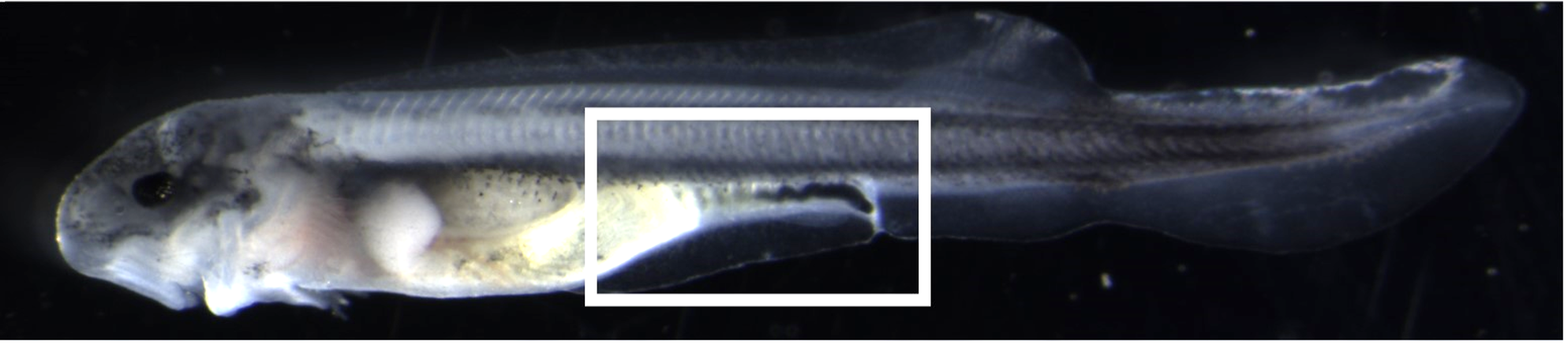
In addition to sturgeons, Jan Štundl also studies a representative of the present-day jawless fishes, the sea lamprey. In a recent study published in Nature (Edens et al., 2024), Prof. Marianne Bronner's team focused on the sympathetic nervous system, which was thought to be completely absent in lampreys for more than a century. The authors were able to show that neural bar cells give rise to the precursors of sympathetic neurons. The scientists obtained these surprising results by studying the late larval development of the lamprey because, unlike other vertebrates, sympathetic neurons develop much later.

Research on lampreys has also contributed to more than a century of debate about how our limbs came to be. An international team from various universities around the world, including Jan Štundl and Prof. Bronner, also contributed to this question (Tzung et al., 2023). The authors focused on very special cells, fibroblasts, found in the preanal region of the fin cuticle, and used several different vertebrate groups, including lampreys, to describe their developmental program responsible for the origin of paired fins.

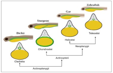
Thus, the aforementioned papers nicely illustrate how research on non-traditional species, including sturgeons from the FROV JU or ectoparasitic sea lampreys, is fundamentally advancing our understanding of vertebrate evolution, as well as the ability to regenerate damaged tissues such as cardiac muscle.

Photo by: Filip Novotný

More information:

A median fin derived from the lateral plate mesoderm and the origin of paired fins

Keh-Weei Tzung, Robert L. Lalonde, Karin D. Prummel, Harsha Mahabaleshwar, Hannah R. Moran, Jan Stundl, Amanda N. Cass, Yao Le, Robert Lea, Karel Dorey, Monika J. Tomecka, Changqing Zhang, Eline C. Brombacher, William T. White, Henry H. Roehl, Frank J. Tulenko, Christoph Winkler, Peter D. Currie, Enrique Amaya, Marcus C. Davis, Marianne E. Bronner, Christian Mosimann & Tom J. Carney . Nature volume 618, pages543–549 (2023). 

Ancient vertebrate dermal armor evolved from trunk neural crest

Stundl, J., Martik, M. L., Chen, D., Raja, D. A., Franěk, R., Pospisilova, A., Pšenička, M.,Metscher, B. D., Braasch, I., Haitina, T., Cerny, R., Ahlberg, P. E., & Bronner, M. E. (2023). Ancient vertebrate dermal armor evolved from trunk neural crest. Proceedings of the National Academy of Sciences, 120(30), e2221120120. https://doi.org/10.1073/pnas.2221120120 (IF=12.779, AIS=4.658).

Neural crest origin of sympathetic neurons at the dawn of vertebrates

Brittany M. Edens, Jan Stundl, Hugo A. Urrutia & Marianne E. Bronner. Nature volume 629, pages121–126 (2024). 

Role of the cardiac neural crest cells in heart development and regeneration

CORDIS, EU research results. 


Evo-devo výzkum na FROV, jehož výsledky byly publikovány v prestižních časopisech

Na konci loňského roku byl na naší fakultě úspěšně ukončen evropský projekt, jehož hlavním řešitelem byl Jan Štundl, který v současné době působí na Kalifornském technologickém institutu ve Spojených státech a nadále úzce spolupracuje s Fakultou rybářství a ochrany vod JU.

Janu Štundlovi se v roce 2020 podařilo získat prestižní grant Marie Skłodowska-Curie, který podpořil jeho postdoktorský pobyt v laboratoři přední světové odbornice na neurální lištu a evolučně vývojovou biologii (evo-devo) prof. Marianne Bronner. Neurální lišta je unikátní buněčná populace, která vzniká během raného embryonálního vývoje. Tyto buňky posléze migrují po celém embryu a dávají vzniknout mnoha rozličným strukturám, mezi něž patří ozubené čelisti, rozličné neurony nebo dentoskeletální elementy, jako jsou šupiny žraloků nebo štítky jeseterů. „Neurální lišta se vyskytuje pouze u obratlovců a vděčíme jí za to, že jsme z evolučního hlediska úspěšným druhem,“ vysvětluje Jan Štundl. Buňky neurální lišty se podílí na téměř každé části našeho těla a hrají navíc významnou roli v procesech regenerace.

V rámci projektu Heart2019 se Jan Štundl zaměřil na zkoumání specifické subpopulace neurální lišty – srdeční neurální lišty. Její buňky během raného vývoje migrují do srdce a hrají klíčovou roli při tvorbě srdečního svalu. Tento projekt navazoval na průkopnický výzkum prof. Marianne Bronner, který naznačoval, že tyto buňky by mohly být zodpovědné za regeneraci srdce. „Na základě výzkumu modelového druhu, zebřičky pruhované (Danio rerio), v laboratoři zjistili, že buňky srdeční neurální lišty skutečně přispívají k regeneraci srdečního svalu,“ upřesňuje Jan Štundl. „Položili jsme si tedy otázku, zdali je tento regenerační fenomén jedinečný pro zebřičky, nebo zda je společný i jiným obratlovcům.“ K jejímu zodpovězení byly zkoumány tři druhy z evolučně významných linií obratlovců – jeseteři, mihule a mloci. Zkoumání mimo jiné potvrdilo, že buňky neurální lišty se podílí na regeneraci srdce také u jiných skupin obratlovců a že můžou být navíc odpovědné také za obnovu dalších tělesných tkání.

V průběhu řešení projektu Heart2019 se navíc podařilo získat celou řadu unikátních výsledků, které se těší mimořádnému zájmu a vyšly v renomovaných vědeckých časopisech, jako je Nature nebo PNAS (Proceedings of the National Academy of Sciences). Jeseteři z FROV JU například přispěli k odhalení původu primitivního pancíře prvních obratlovců a výsledky výzkumu současně poukázaly na fakt, že i buňky neurální lišty mohou produkovat kost mimo hlavu, což zcela změnilo současný pohled na vývojové schopnosti neurální lišty. Tento první „obratlovčí“ pancíř byl složený z kostí a dentinu, látky, která je důležitou součástí našich zubů. Díky této evoluční novince, která vedla ke vzniku a diverzifikaci kožního pancíře podél celé osy těla, naši předci získali ochranu před tehdejšími dravci (Stundl et al., 2023).

Vedle jeseterů studuje Jan Štundl také zástupce současných bezčelistnatců, mihule mořské. V nedávné studii publikované v Nature (Edens et al., 2024) se tým prof. Marianne Bronner zaměřil na sympatický nervový systém, o němž se více jak století předpokládalo, že u mihulí zcela chybí. Autorům se podařilo prokázat, že buňky neurální lišty dávají vzniknout prekurzorům sympatických neuronů. Tyto překvapivé výsledky vědci získali díky studiu pozdního larválního vývoje mihulí, protože narozdíl od ostatních obratlovců se sympatické neurony vyvíjejí výrazně později.

Výzkum na mihulích taktéž přispěl k více jak stoleté debatě o tom, jak vznikly naše končetiny. K této otázce přispěl i mezinárodní tým z různých světových univerzit, jehož součástí byl i Jan Štundl a prof. Bronner (Tzung et et al., 2023). Autoři se zaměřili na velmi zvláštní buňky, fibroblasty, nalézající se v preanální oblasti ploutevního lemu, a na několika různých skupinách obratlovců včetně mihulí popsali jejich vývojový program zodpovědný za vznik párových ploutví.

Výše zmíněné práce tak hezky ilustrují, kterak výzkum netradičních druhů, mezi které patří jeseteři z FROV JU nebo ektoparazitické mihule mořské, zásadním způsobem posouvají naše chápaní evoluce obratlovců, ale i schopností regenerovat poškozené tkáně, jakými jsou například srdeční svalovina.

Foto: Filip Novotný

Pro více informací:

A median fin derived from the lateral plate mesoderm and the origin of paired fins

Keh-Weei Tzung, Robert L. Lalonde, Karin D. Prummel, Harsha Mahabaleshwar, Hannah R. Moran, Jan Stundl, Amanda N. Cass, Yao Le, Robert Lea, Karel Dorey, Monika J. Tomecka, Changqing Zhang, Eline C. Brombacher, William T. White, Henry H. Roehl, Frank J. Tulenko, Christoph Winkler, Peter D. Currie, Enrique Amaya, Marcus C. Davis, Marianne E. Bronner, Christian Mosimann & Tom J. Carney . Nature volume 618, pages543–549 (2023). 

Ancient vertebrate dermal armor evolved from trunk neural crest

Stundl, J., Martik, M. L., Chen, D., Raja, D. A., Franěk, R., Pospisilova, A., Pšenička, M.,Metscher, B. D., Braasch, I., Haitina, T., Cerny, R., Ahlberg, P. E., & Bronner, M. E. (2023). Ancient vertebrate dermal armor evolved from trunk neural crest. Proceedings of the National Academy of Sciences, 120(30), e2221120120. https://doi.org/10.1073/pnas.2221120120 (IF=12.779, AIS=4.658).

Neural crest origin of sympathetic neurons at the dawn of vertebrates

Brittany M. Edens, Jan Stundl, Hugo A. Urrutia & Marianne E. Bronner. Nature volume 629, pages121–126 (2024). 

Role of the cardiac neural crest cells in heart development and regeneration

CORDIS, EU research results. 


Intracytoplasmic sperm injection – a new method of assisted reproduction in sturgeon

  • Fotogalerie:
    • Foto: , Text k fotce: Intracytoplasmic sperm injection procedure
    • Foto: , Text k fotce: Sturgeon larva resulted after ICSI

Intracytoplasmic sperm injection (ICSI) is a widely used method of assisted reproduction in mammals. However, it is not well established in non-mammalian species. In fish, ICSI has been successfully optimized in only a few model species. In this study, we used ICSI for the first time in critically endangered sturgeons. Using a combination of different species (beluga, sterlet, Siberian and Russian sturgeon), we optimized different types of ICSI. We then evaluated fertilization success and paternity of the resulting embryos and larvae. Using single, freshly collected, non-activated sperm, all combinations of crosses produced normal-shaped, food-accepting fish after ISCI. Molecular analysis showed genome integration of both parents in most of the individuals. It can be said that ICSI technology can be used as an assisted reproduction technology to rescue valuable paternal genomes of sturgeon.

Fatira, E., Havelka, M., Saito, T., Landeira, J., Rodina, M., Gela, D., Pšenička, M., 2022. Intracytoplasmic sperm injection in sturgeon species: A promising reproductive technology of selected genitors. Frontiers in Veterinary Science 9:1054345. doi.org/10.3389/fvets.2022.1054345

Written by: doc. Ing. Martin Pšenička, Ph.D.

Intracytoplazmatická injekce spermie – nová metoda asistované reprodukce u jeseterů

  • Fotogalerie:
    • Foto: , Text k fotce: Postup intracytoplazmatické injekce spermie
    • Foto: , Text k fotce: Larva jesetera vzniklá po ICSI

Intracytoplazmatická injekce spermie (ICSI) je široce používanou metodou asistované reprodukce u savců. U nesavčích druhů však není dobře zavedena. U ryb byla doposud ICSI úspěšně optimalizována jen u několika málo modelových druhů. V této studii jsme poprvé použili ICSI u kriticky ohrožených jeseterů. Za použití kombinace různých druhů (vyza velká, jeseter malý, sibiřský a ruský) jsme provedli optimalizaci různých typů ICSI. Poté jsme hodnotili úspěšnost oplození a paternitu vzniklých embryí a larev. S použitím jednotlivých, čerstvě odebraných, neaktivovaných spermií vznikly po ISCI ve všech kombinacích křížení ryby normálního tvaru přijímající potravu. Molekulární analýza prokázala integraci genomu obou rodičů u většiny jedinců. Lze říci, že technologii ICSI lze využít jako technologii asistované reprodukce k záchraně cenných otcovských genomů jeseterů.

Fatira, E., Havelka, M., Saito, T., Landeira, J., Rodina, M., Gela, D., Pšenička, M., 2022. Intracytoplasmic sperm injection in sturgeon species: A promising reproductive technology of selected genitors. Frontiers in Veterinary Science 9:1054345. doi.org/10.3389/fvets.2022.1054345

Napsal: doc. Ing. Martin Pšenička, Ph.D.

Obří náhradní rodič pro danio pruhované

  • Fotogalerie:
    • Foto:
    • Foto:
    • Foto:

Zárodečné kmenové buňky jsou prekurzory gamet, které lze izolovat a transplantovat mezi fylogeneticky příbuznými druhy. Transplantace těchto buněk představuje významný potenciál v reprodukční biotechnologii, protože umožňuje produkci gamet a potomků dárcovského původu prostřednictvím náhradních rodičů. Zebřičky jsou oblíbeným výzkumným modelem díky svým výhodným vlastnostem a bohatým genomickým zdrojům. Jejich malá velikost však omezuje množství získaných spermií. V našem výzkumu jsme využili techniku náhradní reprodukce ke zvýšení produkce spermií u zebřiček s využitím většího druhu Devario, konkrétně dania malabarského (obrovského), který je třikrát větší. Izolovali jsme spermatogoniální buňky ze zralých samců zebřiček a vnesli je do sterilních larev dania malabarského. Půl roku po transplantaci produkoval příjemce spermie zebřiček s trojnásobným objemem a desetinásobnou koncentrací ve srovnání se zebřičkami. Předložená studie je první svého druhu, která produkuje gamety zebřiček prostřednictvím náhradního rodiče odlišného rodu.

Podrobné informace jsou dostupné v původním článku: Nayak, R., Franěk, R., Šindelka, R., Pšenička, M., 2023. Enhancement of zebrafish sperm production via a large body-sized surrogate with germ cell transplantation. Communications Biology 6: 412. https://doi.org/10.1038/s42003-023-04800-7

Obrázek: Schéma studie.

Napsal: doc. Ing. Martin Pšenička, Ph.D.

Unikátní embryonální vývoj střeva u jeseterů: Trávení začíná ještě před prvním příjmem potravy

  • Fotogalerie:
    • Foto: Larva jesetera malého: Exkrece stráveného žloutku před prvním příjmem potravy , Text k fotce: Larva jesetera malého: Exkrece stráveného žloutku před prvním příjmem potravy
    • Foto: Porovnání vývoje střeva mezi rybami (bichir – bichir, sturgeon – jeseter, gar – kostlín a zebrafish – danio). Žlutou barvou je znázorněn žloutek a zelenou barvou trávicí trubice. , Text k fotce: Porovnání vývoje střeva mezi rybami (bichir – bichir, sturgeon – jeseter, gar – kostlín a zebrafish – danio). Žlutou barvou je znázorněn žloutek a zelenou barvou trávicí trubice.

Jeseteři jsou jedni z nejstarších žijících ryb, které se od ostatních obratlovců odlišují jedinečným způsobem embryonálního vývoje střeva. Zatímco u většiny obratlovců žloutek vajíčka/jikry slouží jako zdroj výživy buď intracelulárně (uvnitř buněk endodermu), nebo extracelulárně přes žloutkový váček, jeseteři používají jinou strategii.

Během vývoje embrya vznikají ve vegetativní hemisféře jikry specializované žloutkové buňky, jejichž jedinou funkcí je zajistit endogenní výživu embrya. Tyto buňky jsou postupně obklopeny vyvíjejícím se střevem, které je začíná trávit ještě před tím, než embryo přijme potravu z vnějšího prostředí. Tato jedinečná schopnost trávit vlastní zdroj výživy přímo uvnitř trávicího traktu již během embryonálního vývoje představuje unikátní adaptaci mezi obratlovci.

Primordiální gonocyty (PGCs), prekurzory zárodečných buněk, se však rovněž tvoří z těchto žloutkových buněk. Co je odlišuje od ostatních žloutkových buněk je přítomnost zárodečné plazmy, která PGC specifikuje a určuje jejich budoucí funkci. Po svém vzniku v oblasti vegetativního pólu jikry se PGC musí přesunout z oblasti určené ke trávení do zárodečné lišty, kde se podílejí na vývoji reprodukčního systému. Tato kombinace raného trávení endogenní výživy a současné migrace specifikovaných zárodečných buněk ukazuje na jedinečnost vývojových procesů u jeseterů a jejich unikátní odlišnost od ostatních obratlovců.

Tento objev zdůrazňuje jedinečné přizpůsobení jeseterů, které jim umožňuje optimalizovat výživu a vývoj reprodukčních buněk v embryonálním stadiu. Ukazuje, jak odlišné evoluční strategie přispívají k přežití těchto starobylých rybích druhů. Jeseteři tak nejen potvrzují svou výjimečnost mezi rybami, ale přinášejí i nové poznatky o rozmanitosti vývojových procesů mezi obratlovci.

Podrobné informace lze nalézt v původním vědeckém článku: Shah, M.A., Xie, X., Rodina, M., Štundl, J., Braasch, I., Šindelka, R., Rzepkowska, M., Saito, T., Pšenička, M., 2024. Sturgeon gut development: a unique yolk utilization strategy among vertebrates. Frontiers in Cell and Developmental Biology 12: 1358702. https://doi.org/10.3389/fcell.2024.1358702

Autor: doc. Martin Pšenička

Obr. 1: Larva jesetera malého: Exkrece stráveného žloutku před prvním příjmem potravy

Obr. 2: Porovnání vývoje střeva mezi rybami (bichir – bichir, sturgeon – jeseter, gar – kostlín a zebrafish – danio).Žlutou barvou je znázorněn žloutek a zelenou barvou trávicí trubice. 

Unique Gut Development in Sturgeons: Digestion Begins Before First Feeding

  • Fotogalerie:
    • Foto: Larva of Sterlet Sturgeon: Excretion of Digested Yolk Before First Feeding , Text k fotce: Larva of Sterlet Sturgeon: Excretion of Digested Yolk Before First Feeding
    • Foto: Comparison of Gut Development in Fish. The yolk is shown in yellow and the digestive tube in green. , Text k fotce: Comparison of Gut Development in Fish. The yolk is shown in yellow and the digestive tube in green.

Sturgeons, one of the oldest living fish species, differ from other vertebrates with their unique gut development. While in most vertebrates the yolk of the egg serves as a source of nutrition either intracellularly (within endoderm cells) or extracellularly via the yolk sac, sturgeons have evolved a different strategy.

During embryonic development, specialized yolk cells form in the vegetal hemisphere of the embryo. These cells serve exclusively to provide endogenous nutrition to the developing embryo. As the sturgeon’s gut develops, it gradually surrounds these yolk cells and begins digesting them before the embryo consumes any external food. This remarkable ability to digest its own endogenous nutritional source within the gut during embryonic development represents a unique adaptation among vertebrates.

Primordial germ cells (PGCs), the precursors of reproductive cells, also arise from these yolk cells. What sets them apart from other yolk cells is the presence of germ plasm, which specifies their future role. After their formation in the vegetal pole of the egg, PGCs must migrate away from the digestion zone to the germinal ridge, where they contribute to the development of the reproductive system. This combination of early digestion of endogenous nutrition and the migration of specified germ cells highlights the evolutionary distinctiveness of sturgeon development compared to other vertebrates.

This discovery underscores the sturgeon’s unique adaptations, allowing them to optimize both nutrition and the development of reproductive cells during embryogenesis. It also demonstrates how different evolutionary strategies contribute to the survival of ancient fish species. Sturgeons not only reaffirm their exceptional place among fish but also provide new insights into the diversity of developmental processes in vertebrates.

Detailed information can be found in the original article: Shah, M.A., Xie, X., Rodina, M., Štundl, J., Braasch, I., Šindelka, R., Rzepkowska, M., Saito, T., Pšenička, M., 2024. Sturgeon gut development: a unique yolk utilization strategy among vertebrates. Frontiers in Cell and Developmental Biology 12: 1358702. https://doi.org/10.3389/fcell.2024.1358702

Written by: Martin Pšenička, PhD.

Fig. 1: Larva of Sterlet Sturgeon: Excretion of Digested Yolk Before First Feeding

Fig. 2: Comparison of Gut Development in Fish. The yolk is shown in yellow and the digestive tube in green.

Zůstaňme v kontaktu na
sociálních sítích

Zátiší 728/II, 389 25 Vodňany Tel.+420 387 774 616 | Tato e-mailová adresa je chráněna před spamboty. Pro její zobrazení musíte mít povolen Javascript.

Branišovská 1645/31a, 370 05 České BudějoviceTel. +420 387 774 601 | Tato e-mailová adresa je chráněna před spamboty. Pro její zobrazení musíte mít povolen Javascript.

© Jihočeská univerzita v Českých Budějovicích
Cookies

1